Posts

Showing posts from 2018

Med Legal Art - Disc Herniation Boards

Image
These illustrations are colored interpretations of cervical and lumbar disc herniations from MRI films for 2 different patients (or plaintiffs). The layouts include flat-graphic orientation images to give viewers familiarity with the level of the scans, the original MRI (cropped to the injury), and digital paintings to highlight the areas of interest.

Glucose-stimulated Insulin Secretion

Image
This molecular illustration shows the components involved in the release of insulin from a pancreatic beta-cell during a period of elevated glucose-levels. The insulin molecules will go on to bind to insulin-receptors on other cells that allow glucose to enter and be used for energy.

Medial View of Raccoon Mandible

Image
This image is half of a raccoon mandible that was recovered in marshes along the Georgia coast. Nerve innervation and arterial supply for the teeth can be seen entering through the mandibular foramen. A section of the body has been removed to reveal the structures in the mandibular canal. The sample is missing the incisors.

Membrane-Attack Complex (MAC)

Image
This molecular illustration shows the steps involved in building the ‘membrane-attack complex’ (MAC) during an immune response against an invading bacterium. A transmembrane channel is formed when up to 16 C9 molecules are added, thus disrupting the outer membrane (the complete pore was halved for the sake of the illustration).

Bovine Cervical Vertebra Anatomy (revisited)

Image
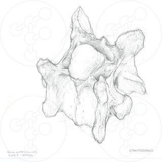
These images are of a bovine (cow or ox) cervical vertebra that was found in marshes along the Georgia coast. The transverse foramen and short spinous process are key anatomical identifiers for the group of cervical vertebrae. One image shows the bone by itself and the other shows the spinal cord in place; structures in both images are labeled for reference.

Bovine Cervical Vertebra

Image
This is a sketch of a bovine (cow or ox) cervical vertebra that was found in marshes along the Georgia coast. The transverse foramen ( vertebrarterial canals ) and short spine are key anatomical identifiers for the group of cervical vertebrae. ( Unfortunately, the bone is ‘standing up’ on the posterior end and needs to be redrawn in the bone’s true orientation [lying down]. This is what happens when someone trained in drawing HUMAN vertebrae draws a mammalian tetrapod’s vertebra. Oops… It’ll be redrawn soon. )

Ancient Coyote Skull

Image
The original sketch for this infographic was made at a ‘SciArt Meetup’ event at the Florida Museum of Natural History (color and graphical details added afterwards). The graphics on the left include a breakdown of the time period and geographical location of the ancient animal’s existence.

Conotoxin

Image
This is an image of conotoxin and an inset of how it affects a voltage-gated calcium channel (VGCC). Conotoxin is a peptide toxin created by a tropical marine snail ( Conus victoriae ). Aside from VGCCs, the toxin also targets voltage-gated sodium channels, voltage-gated potassium channels, and nicotinic acetylcholine receptors. Conotoxins are being explored as an option for pain-relief therapy.

Figure Drawing

Image
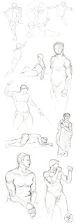
Some sketches from a figure drawing session at a local art museum; pose durations were between 2 and 30 minutes.

Figure Drawing

Image
Some sketches from a figure drawing session at a local art museum; pose durations were between 2 and 30 minutes.

AJC Peachtree Road Race 2018 - logo design entry

Image
This design was created and submitted for consideration as the AJC Peachtree Road Race 2018 logo. The design was inspired by 80’s-styled movie posters. A silhouette of the city is back-lit by a setting sun (in the shape of a peach). The typeface and colors follow the reference design-pattern. This style was chosen for it’s relaxed vibe and fun color palette.

Product Design Sketch for a Presentation

Image
This sketch was commissioned to be part of a product design presentation. The new product will work in conjunction with the Apple TV device. This sketch shows the Apple TV device in an interesting perspective, with ‘blueprint-like’ lines and measurements surrounding it, to reinforce the idea that this is part of a bigger, conceptual project. The original image was scaled to fit a widescreen Microsoft Powerpoint slide.

Memory Depotentiation at the Synapse

Image
This image generalizes the electrochemical steps involved in memory depotentiation at the synapse between neurons. A stimulus activates NMDAR, which then permit calcium entry into the cell. As the cell depolarizes, VGCC activates and allows more calcium to flow into the cell. This intracellular calcium-oscillation mobilizes proteins that will eventually cause the cell to degrade the AMPA receptors by means of endocytosis. * This is a variation of an image I created for the article “Harnessing Electroceuticals to Treat Disorders Arising from Traumatic Stress: Theoretical Considerations Using a Psychosensory Model” in “Explore: The Journal of Science and Healing” .

Chlorpyrifos

Image
This image, sized for a science journal, shows one effect of chlorpyrifos on the human body, particularly in the nervous system. In the ‘normal’ scenario, the enzyme acetylcholinesterase (or cholinesterase) breaks down the neurotransmitter, acetylcholine. In the clinical scenario, the pesticide inhibits the enzyme from working properly, thus leading to an accumulation of acetylcholine. The build-up can cause over-stimulation of nerves, muscles, and glands.