Posts

Med Legal Art - multiple injuries and corrections

Image
These medical legal boards feature colored medical films of numerous herniations and surgeries for a single personal injury case. Each layout includes a flat-graphic orientation image to give jurors familiarity with the area being scanned or operated on. The medical film boards have the original medical film and a colored interpretation of the film; the surgical boards display the steps involved in correcting the injury.

Molecular Pathway for Virus Entry

Image
This molecular illustration shows the membrane-bound proteins involved in the uptake of a virus particle into the cell. Reading from left to right, the arrows show how the proteins pass the virus down the line until it reaches the final protein that will trigger the endocytosis.

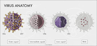
Rotavirus Anatomy

Image
This scientific illustration inset (from a larger poster) shows the anatomy of a rotavirus particle. The different capsids that surround the RNA strands are isolated and positioned within a flat, gray virus shape to show their relevant size and position within the complete virus.

Med Legal Art - Disc Herniation Boards

Image
These illustrations are colored interpretations of cervical and lumbar disc herniations from MRI films for 2 different patients (or plaintiffs). The layouts include flat-graphic orientation images to give viewers familiarity with the level of the scans, the original MRI (cropped to the injury), and digital paintings to highlight the areas of interest.

Glucose-stimulated Insulin Secretion

Image
This molecular illustration shows the components involved in the release of insulin from a pancreatic beta-cell during a period of elevated glucose-levels. The insulin molecules will go on to bind to insulin-receptors on other cells that allow glucose to enter and be used for energy.

Medial View of Raccoon Mandible

Image
This image is half of a raccoon mandible that was recovered in marshes along the Georgia coast. Nerve innervation and arterial supply for the teeth can be seen entering through the mandibular foramen. A section of the body has been removed to reveal the structures in the mandibular canal. The sample is missing the incisors.

Membrane-Attack Complex (MAC)

Image
This molecular illustration shows the steps involved in building the ‘membrane-attack complex’ (MAC) during an immune response against an invading bacterium. A transmembrane channel is formed when up to 16 C9 molecules are added, thus disrupting the outer membrane (the complete pore was halved for the sake of the illustration).